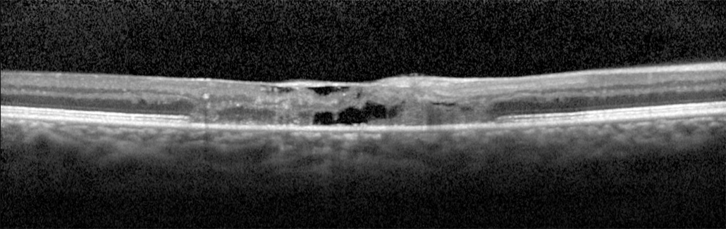
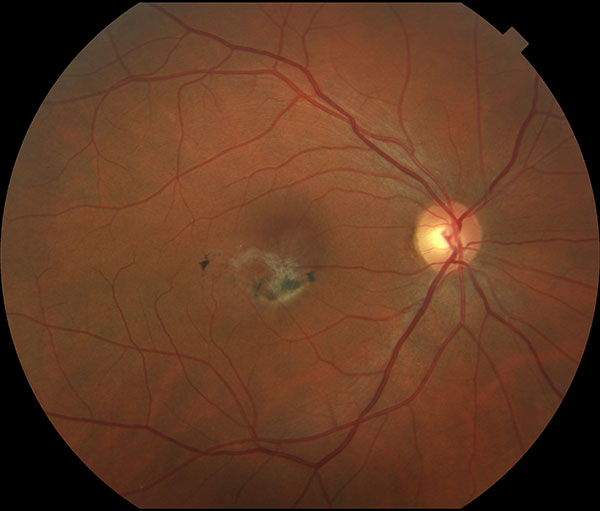
Idiopathic Perifoveal Telangiectasis is commonly referred to as macular telangiectasia, a retinal disorder that affects the center most part of the macula. This poorly understood disease is most common in people between 40 to 60 years old. Dilated and leaky retinal capillaries develop around the temporal part of the foveal area and may eventually encompass the entire area, causing pigmentation to migrate below the retina and into the vitreoretinal interface. As this progresses, visual clarity decreases.
Dr. Freund is everything one would want in a doctor: conscientious, ethical, knowledgeable, and experienced. He is ready, able and willing to explain to you your diagnosis and treatment. He’s also very personable. Highly recommended.
JOHN G. GoogleThe term parafoveal telangiectasia has a wide variety of names, including macular telangiectasia, idiopathic parafoveal telangiectasia, idiopathic perifoveal telangiectasia, perifoveal telangiectasia, juxtafoveal retinal telangiectasia, telangiectasis, and MacTel. The condition is quite rare and impacts the small blood vessels in the retina, leading to abnormal dilation (enlargement).
Parafoveal telangiectasia is usually divided into a few types, with different blood vessel changes for each. Based on Dr. Yannuzzi’s study, which reviewed the frequency and nature of idiopathic macular telangiectasia and classified the disorders based on new clinical and imaging observations, there are 2 distinct types of this condition, including:
There is also a third type of macular telangiectasia called occlusive telangiectasia which is extremely rare and poorly understood. It is characterized by progressive visual loss with more diseased vessels and occlusion of the capillaries of the fovea in both eyes.
The symptoms of Macular Telangiectasia can vary depending on the stage and severity of the condition. Early stages may not involve any symptoms or lead to only mild visual changes.
Symptoms you may develop as the condition progresses include:
If you are experiencing any of these symptoms, it is important to see a telangiectasia doctor for a comprehensive exam to slow the progression of the disease and preserve your vision.
Parafoveal Telangiectasia is a rare retinal disorder, and its exact cause is not fully understood.
However, some factors believed to result in the condition include:
Though there is no effective cure for macular telangiectasia at the moment, your doctor can help control symptoms and prevent further vision loss.
Some of the treatments the specialist may recommend include:
As the best telangiectasia specialist in NYC and the entire country, Dr. Yannuzzi can precisely determine the stage and severity of your condition and find the most appropriate treatment options to preserve your eyesight. Contact VRMNY and achieve healthy eyes.
Let us help you enjoy your life
Call: (212) 861-9797To Speak With An Appointment Coordinator Now